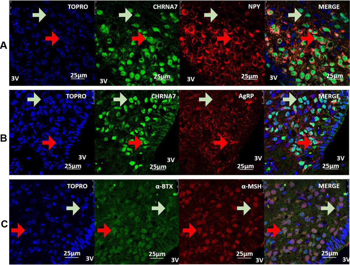
Fig. 1. AchRα7 and NPY/AgRP/α-MSH neurons are colocalised in the hypothalamus. Confocal images illustrating (A) NPY neurons (red) and AchRα7 (green), (B) AgRP neurons (red) and AchRα7 (green), (C) α-MSH neurons (red) and αBgt (green); nuclear labelling was done with TO-PRO3 (blue) and images were merged. Staining was performed on coronal sections of the brain (adult male mouse, 7 weeks old). 1000x magnification. V3, third ventricle (n=4).